What is DSAEK?
Descemet’s Stripping Automated Endothelial Keratoplasty is a surgical treatment for people suffering a corneal endothelial dysfunction.
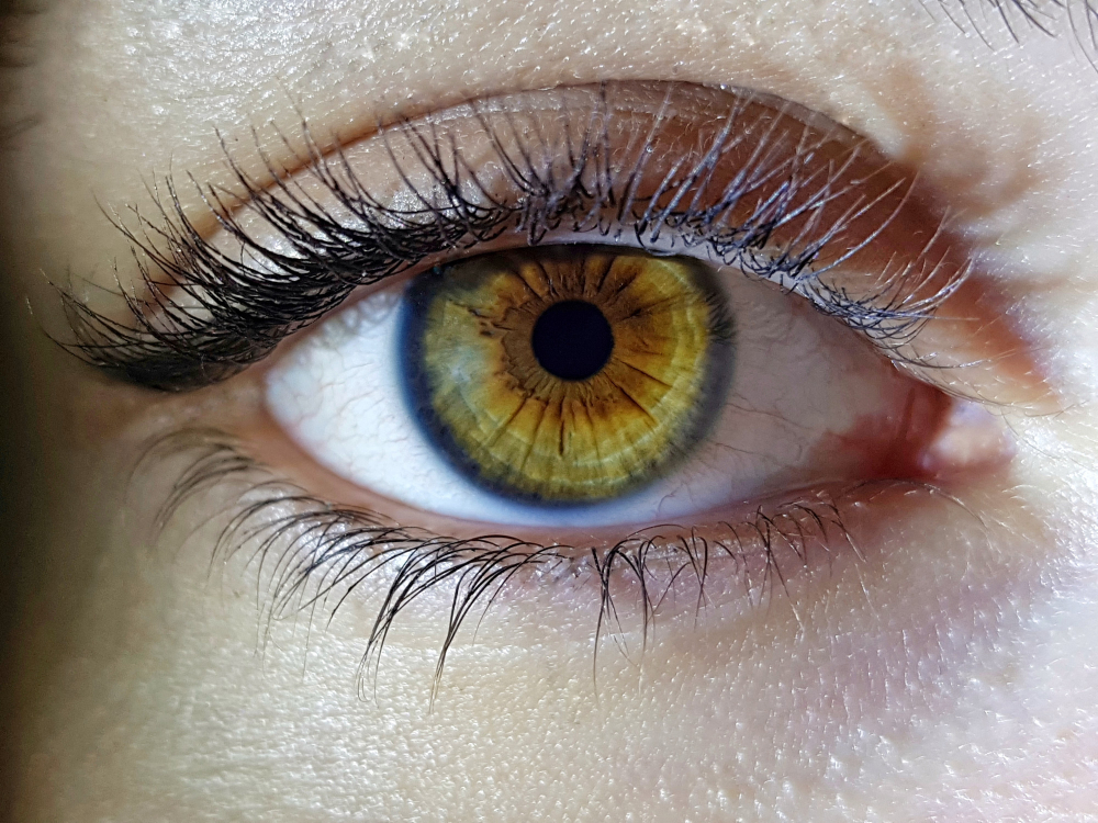
What is the cornea?
The cornea is often described as a clear window located at the front of the eye. The inner layer of the cornea is covered by the endothelium, a thin layer of cells. The main goal of the endothelium is to extract water out of the cornea. This mechanism helps it stay clear.
Endothelial cells are an essential part of the cornea : if they are damaged, an oedema appears and the cornea becomes cloudy, which makes the vision blurry[1].
What causes endothelial cells damage?
Most cases of endothelial cells damage are caused by a disease called “corneal dystrophy of Fuch’s”. Endothelial cells can also be lost after an eye injury or a trauma[2].
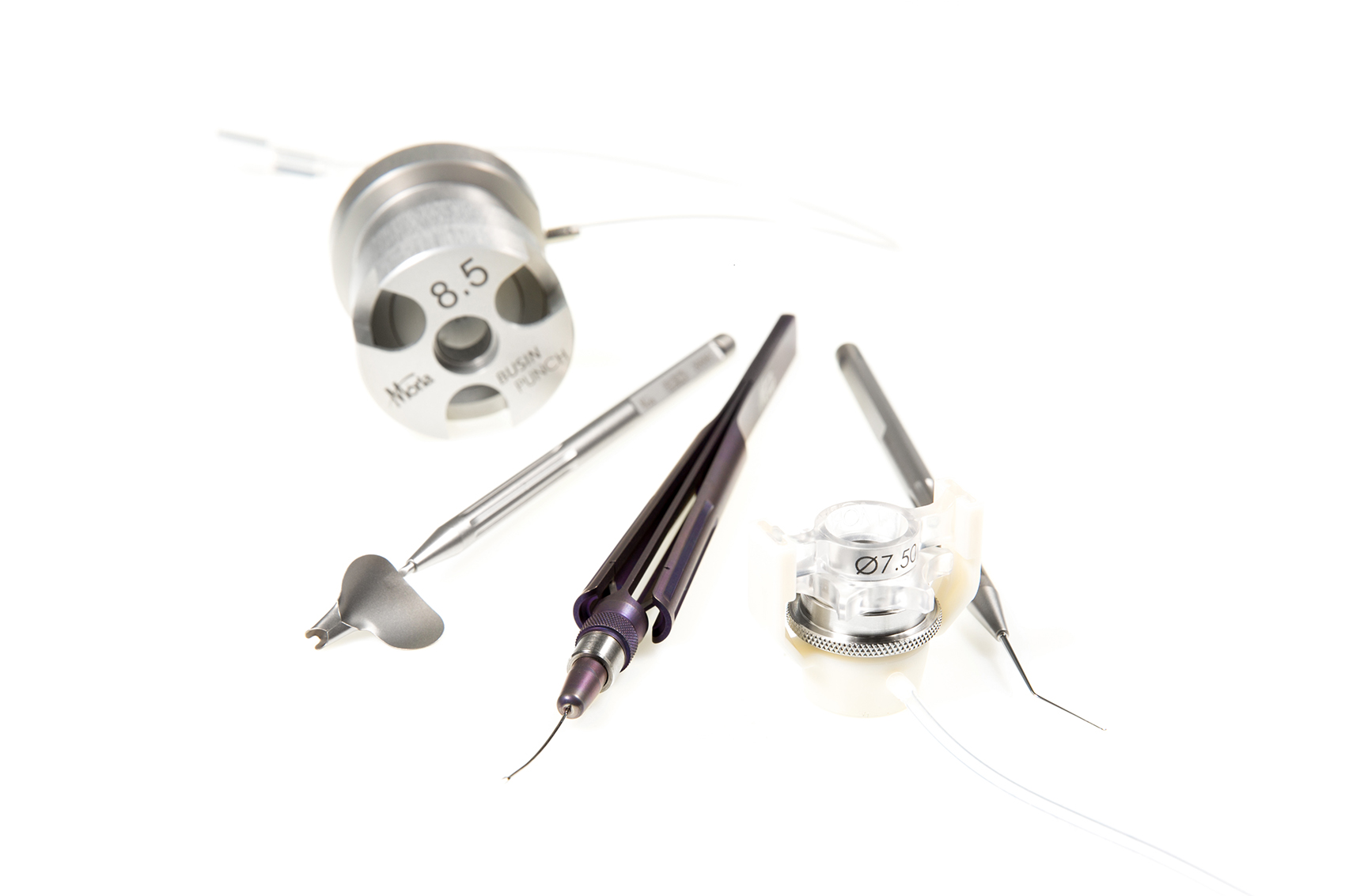
Instruments needed for DSAEK
DSAEK is a surgical intervention that requires a high level of precision. For this purpose, Moria offers a wide range of DSAEK dedicated tools that were designed to help ophthalmic surgeons achieve success during the operation:
[1] Keith M. Meek∗ and Carlo Knupp - Corneal structure and transparency - Pubmed Central - https://www.ncbi.nlm.nih.gov/pmc/articles/PMC4655862/
[2] Hussain Elhalis, MD,1,3,* Behrooz Azizi, MD,1,3,* and Ula V. Jurkunas, MD1,2,3 - Fuchs Endothelial Corneal Dystrophy - Pubmed Central - https://www.ncbi.nlm.nih.gov/pmc/articles/PMC3061348/
In which cases is DSAEK required?
DSAEK is usually successful in people at an early stage of a corneal endothelial disease, when the oedema is at its first stage and the vision is not completely blurry. In case the corneal endothelial disease is more serious, then there is a risk of stromal scarring remaining after the operation. The patient will thus not recover completely[1].
What has to be checked prior to DSAEK?
Before performing DSAEK, the eye surgeon has to verify the stability of the eye and to make sure there is no active macular edema. In addition, the intraocular pressure must not be elevated. Other conditions have to be checked as well, as they might make the intervention more difficult. These are mainly glaucoma, the presence of a filtering bleb, the presence of a narrow anterior chamber (that would not be deep enough) and the presence of an anterior chamber phakic intraocular lens.
[1] Sepehr Feizi - Corneal endothelial cell dysfunction: etiologies and management - Pubmed Central - https://www.ncbi.nlm.nih.gov/pmc/articles/PMC6293368/
Are there any risks associated with DSAEK?
The condition of some categories of persons increases the risks of complications, regardless of how well the intervention has been performed. This is for example with people who suffer dementia as they might rub the operated eye, which can result in a graft detachment. Also, patients with glaucoma who have a trabeculectomy filtration are at higher risk from postoperative hypotony and graft dislocation[1].
[1] Annie Stuart - Performing DSAEK: A Step-by-Step Guide - American Academy of Ophthalmology - https://www.aao.org/eyenet/article/performing-dsaek-stepbystep-guide
How is DSAEK performed?
DSAEK is performed in four main stages :
Wound size
Surgeons make a limbal wound in order to reduce endothelial cell loss from the donor’s insertion using Busin spatula and Busin forceps by Moria. This size of wound makes the insertion safer.
Air bubble
The most important part of the adhesion of the corneal donor is the air bubble tension. If this point is not thoroughly controlled, it can lead to pupillary block and eye vision damage.
To avoid this, the air bubble is created within the anterior chamber, left for an hour, then the bubble is burped at the slit lamp until the bottom of the pupillary edge is cleared. The patient must not bend over for 24 hours to avoid a migration of the residual bubble behind the pupil.
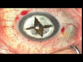
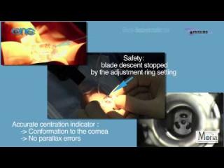
Centration
The donor’s cornea surgical centration must be done in a way to cover the pupil with no angle touch.
Rebubbling
Donor dislocations may occur. Although they are not considered as an emergency, most surgeons prefer to treat this quickly. If it is correctly done, rebubbling doesn’t affect the clarity of the graft[6].
[6] MARK S. GOROVOY, MD - Maximizing the Results of DSAEK - Cataract and Refractive Surgery today - https://crstoday.com/articles/2011-apr/maximizing-the-results-of-dsaek/
What are the postoperative instructions?
During the hour that follows DSAEK, the patient lies in the recovery room. Surgeon then examines patient to ensure the graft is properly positioned and in the right way. If this is not the case, the patient will be taken back to the operating room where another air bubble is inserted to reposition the graft.
At home
Patients are asked to lie on their back for at least 24 hours. Eating and bathing are the only exceptions until the first follow-up consultation with the eye surgeon. If there’s any pain or discomfort, the Doctor has to be notified quickly as this could be a sign of pupillary block.
Medication to be taken
Antibiotics and topical steroids will be started on the day of the surgery or on the next day, depending on the Doctor’s instructions. They are usually taken four times a day with cycloplegics to be taken three times a day. As long as the air bubble is present, cycloplegics have to be continued. Antibiotics are usually taken for ten days while steroids must be taken from one to six weeks, according to the ophthalmic surgeon’s prescription[7].
Follow-up consultations
The first follow-up visit will take place on the day that follows the intervention. There will be another visit in the following month, then after three months.
During the first visit, the eye surgeon will check the remaining air bubbles. If there is a detachment, the graft will be repositioned.
[7] Annie Stuart - Performing DSAEK: A Step-by-Step Guide - American Academy of Ophthalmology - https://www.aao.org/eyenet/article/performing-dsaek-stepbystep-guide
Possible side-effects after DSAEK surgery
Possible complications following DSAEK are :
- endothelial rejection (in very rare cases)
- graft dislocation
- air-induced pupillary block
- bacterial endophthalmitis (in very rare cases)
- secondary glaucoma
- secondary graft failure in the late postoperative period[8]
[8] Samar K Basak and Soham Basak1 - Complications and management in Descemet's stripping endothelial keratoplasty: Analysis of consecutive 430 cases - National Library of Medicine - https://www.ncbi.nlm.nih.gov/pmc/articles/PMC4005239/
DSAEK advantages
DSAEK is considered by surgeons as being a less-invasive surgery compared to full-thickness keratoplasty. Although it involves some surgical challenges, it offers more benefits than standard penetrating keratoplasty.
Most patients and surgeons show satisfaction about the visual outcomes of this DSAEK procedure.
80% of the patients can see their vision corrected by 6 weeks while this figure increases to 90% by three months