How is cataract surgery performed?
What are the cataract surgery steps with instruments ?
Anesthesia
Before starting the intervention, the ophthalmologist applies an anesthetic agent in the shape of eye drops or injection. The injection can be retrobulbar (behind the globe of the eye) using a retrobulbar needle or peribulbar (above and below the orbit)[1].
Incision
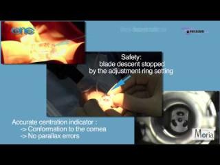
Once the anesthesia is completed, an incision is done. In most cases, the incision is made in the cornea as it shows many advantages : it needs less anesthesia, it allows a fast recovery and it is self-sealing, it gives the surgeon an easier access to the anterior chamber, the risks of bleeding are lower than with a scleral tunnel incision[2].
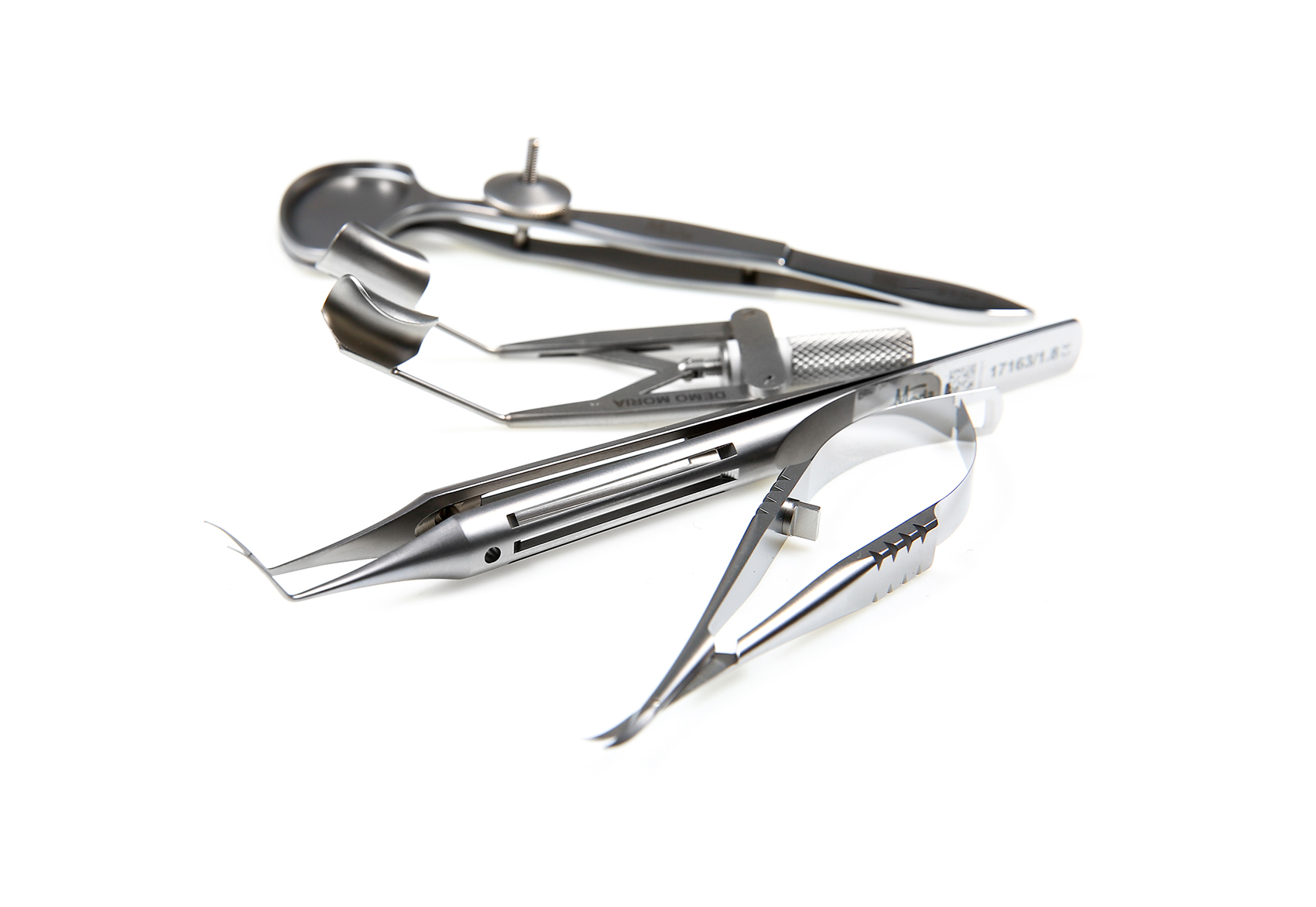
The incision is made using a blade, then a Haefliger Phaco Cleaver as example is used to prevent damaging the posterior capsule during phacoemulsification.
Capsulorhexis
Capsulorhexis, also called “continuous curvilinear capsulorhexis”, ensures the success of phacoemulsification. This procedure is performed by the surgeon during cataract surgery in order to remove the lens capsule from the eye using capsulorhexis forceps. It is done to create a circular opening in front of the bag that contains the cataract[3].
Nuclear disassembly and aspiration
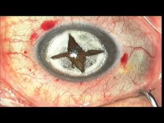
Using ultrasounds, the lens is fractionated into four quadrants and each one is then aspirated. Using lateral pressure, the posterior plate and the nuclear rim are fractured. This step is performed using two instruments: a probe and a spatula.
IOL implantation
The surgeon must make sure that the capsular bag is empty. Any remaining fragment could lead to a postoperative infection. At this stage, the intraocular lens is implanted within the empty capsular bag. Use of specific instruments, such as the IOL haptic Gorovoy marker, helps to correctly position specific IOLs[4].
[1] ROGER F. STEINERT, MD - The basics of phacoemulsification - Cataract and refractive surgery today (En ligne) - https://crstoday.com/articles/2011-may/the-basics-of-phacoemulsification/#:~:text=The%20key%20surgical%20steps%20are,8)%20closure%20of%20the%20incision.
[2] Benedito Antônio de Sousa1, Anderson Teixeira1-3*, Camila Salaroli2, Nonato Souza2 and Lucy Gomes4 - Wound architectural analysis of 1.8mm microincision cataract surgery using spectral domain OCT - HSPC (En ligne) - https://www.clinophthaljournal.com/articles/ijceo-aid1020.pdf
[3]Cataract surgery steps: from start to finish - Vision and Eye Health ( En ligne ) - https://www.vision-and-eye-health.com/cataractsurgery-steps.html
[4] Kierstan Boyd - IOL Implants: Lens Replacement After Cataracts - American Academy of Ophthalmology (En ligne) - https://www.aao.org/eye-health/diseases/cataracts-iol-implants
How to choose the best cataract surgery equipment?
Cataract surgery needs to be performed by experienced surgeons, using quality equipment. For a successful surgery, it is important to choose instruments made with the best materials and designs.
Before purchasing cataract surgery instruments, one should consider some important points[1]:
- the need for the instrument
- a careful evaluation of the product via a clear conversation with a member of the company where the product is about to be purchased
- the existence of a technical support service
- whether or not the product is reusable and how often a maintenance must be carried out.
Whether you are looking for instruments designed to perform cataract surgery techniques such as phacoemulsification, stop-and-chop, bimanual and extracapsular, or if you’re willing to purchase cataract surgery sets, Moria instruments have proven to be among the best ones for more than 200 years.
Moria is a French company that was established in France in 1820. Over the years, it has developed a special know-how for everything related to ophthalmic surgery. Moria team is made up of experts in ophthalmology that all together aim to develop instruments that ensure safe and successful surgeries.
